CHIROPRACTIC TREATMENT OF LUMBAR SPINE DISC INJURIES AND HOW THIS DIFFERS FROM CONVENTIONAL THERAPY.

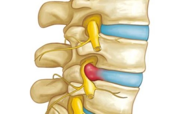
In the last blog we discussed what lumbar spine disc injuries are and how they happen. It all translates back to too much pressure being placed on a particular spinal level(s), causing the disc to prolapse (bulge) outwards resulting in pressure on neural structures.

Conventional therapy focuses on treating the injured level itself with the aim of reducing the size of the disc bulge. This can include rest, analgesics and anti inflammatories. While this approach may yield temporary symptomatic relief it is effectively doing nothing about THE ROOT CAUSE OF THE PROBLEM. More often than not the disc will repeatedly relapse a short while later because the dysfunction has not been corrected. It is effectively like putting a plaster over the problem.

CHIROPRACTIC TREATMENT CORRECTS WHAT IS CAUSING THE DISC TO PROLAPSE:
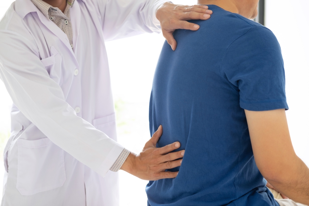
•By correcting and normalizing the spinal biomechanics (movement patterns) throughout the spine, your Chiropractor ensures that all load is distributed equally. Joints which are not moving normally can simply not function normally forcing adjacent levels to carry extra load, loading the discs excessively. Specific Chiropractic manipulation/mobilization corrects this dysfunction.

•Structural anomalies such as excessive scoliosis/kyphosis/lordosis are addressed and corrective exercises are given.

•Leg length inequalities are treated if within the ‘functional difference ‘ category but may be referred for orthotics if structural in nature.

•Muscle strength/flexibility imbalances are addressed and corrective exercises are given.

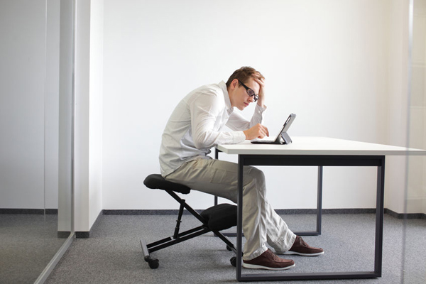
•Postural correction for standing/sitting/sleeping postures is vital and corrective advice regarding occupational/sporting actions invaluable.
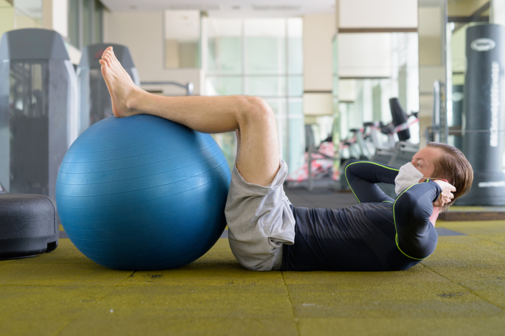
All of the above puts Chiropractic at the forefront for the conservative treatment of lumbar spine disc injuries.The holistic approach prevents future relapses.Your Chiropractor will always refer you when necessary but always try conservative therapy first! Written by Dr. E Greyling

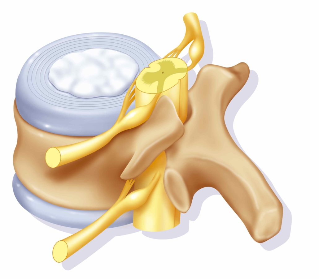
LUMBAR SPINE DISC INJURIES ARE ONE OF THE MOST COMMON SPINAL INJURIES SUFFERED GLOBALLY. IT IS ESTIMATED THAT UP TO 70 PERCENT OF THE WORLDS POPULATION WILL SUFFER SUCH AN INJURY AT SOME STAGE OF THEIR LIVES.

In between the lumbar vertebrae of the spine there are fibro-cartilaginous cushions or discs known as the intervertebral discs. Each disc is comprised of two parts, a tough outer cartilaginous ring known as the annulus fibrosis which encases a soft inner gelatinous core known as the nucleus pulposis.

Occasionally the nucleus pulposis (gelatinous core) will push outwards through the annulus fibrosis due to excessive pressure on that level. This causes the formation of a bulge or prolapse in the outer rim of the disc. If the prolapse is in the direction of the nerve root or spinal cord this can cause pressure on these structures resulting in neurological symptoms.

HOW DOES THIS HAPPEN?
Lumbar disc prolapses happen when there is excessive pressure on a particular spinal level(s). This can occur due to:
•Bad posture
•Bad habits and incorrect sporting actions
•Repetitive lifting injuries
•Weak core musculature
•Spinal anomalies such as scoliosis etc.
•Leg length inequality translating imbalance
up the spine.
•Spinal joint dysfunction etc

WHAT ARE THE SYMPTOMS OF A LUMBAR DISC PROLAPSE?
•Syptoms may /may not include back pain.
•Pain in the buttock or sciatic notch.
•Pain into one or both legs.
•Parasthesia or pins and needles into one or both legs.
•Numbness into one or both legs.
•Weakness into one or both legs usually in a
particular movement.

DO ANY OF THESE SYMPTOMS SOUND FAMILIAR? THEN STAY TUNED FOR NEXT WEEK’S BLOG WHERE WE WILL DISCUSS HOW CHIROPRACTIC TREATMENT OF A DISC INJURY DIFFERS FROM CONVENTIONAL THERAPY AND HOW WE CAN HELP YOU!- Written by DR E. Greyling

DOES IT LOOK OR FEEL AS IF ONE LEG IS LONGER THAN THE OTHER?

If this is indeed you, the most important thing to establish is whether the leg length discrepancy is structural or functional in nature.

Treatment simply cannot resume before the origin of the discrepancy has been established. Basic measurement with a measuring tape is a helpful tool to indicate whether there is a significant leg length difference and whether an X-ray is necessary. An X-ray is the only definitive and diagnostic way to truly measure a leg discrepancy.

STRUCTURAL LEG LENGTH DISCREPANCY implies a physically longer/shorter leg on the one side. This can be quite common post fracture or surgery, especially if the patient was still growing at the time. Hip and knee replacements can also cause this presentation. The important thing to note is that up to 80 percent of the population has a slight structural leg length discrepancy of less than 5mm. A difference of less than a 5mm margin is usually deemed negligible and in most cases WILL NOT WARRANT INSOLES OR HEEL LIFTS.

FUNCTIONAL LEG LENGTH DISCREPANCY implies that there is no (or only marginal) physical difference between leg lengths despite the one leg presenting notably longer. This occurs due to a dipping or a twisting of the pelvis on one side, causing a longer leg presentation. The patient may complain that ‘they look skew in the mirror’,’keep tripping over one foot,’ or ‘feel the need to hike one leg while walking.’ Lying these patients on their backs with legs totally extended and comparing the medial malleoli of the ankles, will often reveal a longer leg on one side. As mentioned, this is not necessarily physically or structurally so, hence, a functional difference.

Functional leg length discrepancies can be caused by a myriad of reasons including:
•Bad habits causing pelvic torsion or twisting of the pelvis such as :
Sitting with the legs crossed,
Standing with all the weight continually on one leg,
Continually sitting on an uneven surface, such as a wallet in the back pocket.
•Incorrect sporting/working actions favouring one side resulting in muscles being stronger around one side of the pelvis.
•Weakness/Inflexibility of the muscles around one side of the pelvis.
•Lumbar spine or sacro-iliac joint dysfunction resulting from bad posture/injury.
•Lumbar spine scoliosis.
•Foot problems such as excessive pronation on one side etc.

Thankfully Chiropractic specializes in identifying , diagnosing and treating the ROOT CAUSE OF THE PROBLEM. Your Chiropractor will go about restoring normal joint function , balancing uneven muscle strength/flexibility and correcting all actions potentially causing the problem. Prescribed exercises and home advice are vital for maintaining the treatment effect.

Every individual case is taken according to its merit -even in patients with significant leg length (over 5ml ) differences. However, if any patient does not show sustainable symptomatic progress or keeps relapsing despite corrective treatment -then it may be time for foot orthotic referral! This involves a slightly raised insole on the short leg side, it is extremely comfortable and the difference is often immediately notable. Larger structural differences over 10 ml are often referred immediately, especially in athletes.

So don’t panic if you suspect one leg to be longer than the other -odds are that it is a functional discrepancy and absolutely treatable! Written by Dr. E GREYLING

DO YOU EXPERIENCE PAIN ON THE OUTSIDE OF THE KNEE WHEN WALKING, RUNNING OR CYCLING? YOU COULD BE SUFFERING FROM ILIOTIBIAL BAND SYNDROME AND HERE IS HOW CHIROPRACTIC CAN HELP YOU.

Iliotibial band syndrome (ITBS) is a friction syndrome of the iliotibial band (ITB) over the lateral femoral condyle of the knee, causing pain and inflammation. The ITB is a tough tendinous band which originates as the gluteus maximus and tensor fascia lata muscles merge from the pelvis. The ITB then runs down the outside of the thigh, crossing the lateral knee over the lateral femoral condyle and attaches to the outside of the tibia (shin bone).

ITBS is classically defined as an overuse injury common in runners and cyclists, but it can occur in sedentary or inactive individuals. ITBS is easily diagnosed because of the obvious signs and history. It is, however, far less correctly treated because of the wide range of variables influencing knee joint biomechanics. These range from spinopelvic alignment issues to muscle imbalances around the hip and knee, through to foot problems.

THE TYPICAL CAUSES OF ITBS RESULT IN INCREASED ITB TENSION AND TORSIONAL PRESSURE ON THE KNEE.THIS INCREASES FRICTION BETWEEN THE ITB AND THE LATERAL FEMORAL CONDYLE. CAUSES INCLUDE (AMONGST OTHERS) :

-Weak lumbar spine muscles causing a slouched posture and compensatory pronation at the feet.
-Pelvic torsion where the pelvis rotates about its axis causing a functional leg length difference.
-Excessively tight gluteus maximus and tensor fascia latae muscles which pulls the ITB too tight at the knee.
-Weak hip abductors including gluteus medius causing the pelvis to dip on the non weight-bearing side while walking/running- this tensions the ITB on the weight-bearing side.
-Foot problems such as pes planus (flat foot) or excessive foot pronation.
-Weak lower limb muscles such as tibialis posterior and calf muscles causing a longer toe-off action when walking or running resulting in compensatory foot pronation.

THANKFULLY CHIROPRACTIC ASESSES, ADRESSES AND TREATS ALL OF THE ABOVE:

Once your Chiropractor has corrected any lumbopelvic alignment issues, they will go about testing the strength and flexibility of related muscles down the leg. Myofascial trigger point therapy, strengthening and stretches are applied specific for each individual’s problem. Running style is assessed and bad habits are corrected. Home exercises are advised and CORRECT rolling of the ITB is especially recommended.

Anyone suffering with/recovering from ITBS should avoid running hills both up and down! Avoid running on a camber on the road and especially avoid very soft grassy tacks/trail running at first.

It is highly recommended that all runners have their foot action assessed by a reputed running store or podiatrist to make sure they are wearing the correct footwear, specific to their walking/running pattern.

If a leg length difference is structural or if foot problems can’t be rectified with exercise, referral to a podiatrist for orthotics may be indicated. YOUR CHIROPRACTOR WILL ALWAYS REFER YOU WHEN INDICATED. Surgery remains a poor option in the treatment of ITBS as it usually just adresses the symptomatic knee and not the actual cause of the problem.
Written by Dr. Etienne Greyling.

DO YOU SUFFER PAIN, NUMBNESS OR PINS AND NEEDLES IN THE HAND? YOU COULD BE EXPERIENCING SYMPTOMS OF CARPAL TUNNEL SYNDROME AND HERE IS HOW CHIROPRACTIC CAN HELP YOU:

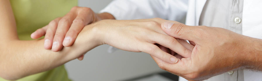
Carpal tunnel syndrome is an impingement of the median nerve at the wrist as it passes through the carpal tunnel (a small passage created between the wrist bones and transverse ligament of the wrist.)

Patients will usually describe the hand as going dead, pain, pins and needles or weakness in grip strength. Symptoms may be aggravated by certain activities but actions as innocuous as an extended wrist position while sleeping may bring them on.

The condition is very often misdiagnosed and to understand this one has to understand the pathway of the median nerve. The nerve originates in the cervical spine (from the medial and lateral cords of the brachial plexus C5-T1). The nerve then courses through the axilla (armpit area) and crosses the elbow through the cubital fossa (elbow crease). It then travels down the arm between the forearm muscles and enters the hand through the carpal tunnel. The median nerve goes on to supply the palmar surface of the thumb, forefinger, middle finger and medial half of the ring finger. Symptoms anywhere other than in this median nerve distribution simply CANNOT be carpal tunnel syndrome.

Entrapment of the median nerve at any of the abovementioned anatomical zones, from the neck through the forearm muscles (pronator muscle especially) to the hand, can reproduce the exact same symptoms as carpal tunnel syndrome. Your Chiropractor will use special orthopedic testing to determine the exact area of nerve entrapment and treat you accordingly.

In true carpal tunnel syndrome the carpal tunnel has been narrowed or compromised as a result of:
-Trauma to the wrist causing misalignment of the carpal bones.
-Shortening of the transverse ligament of the wrist, or inflammation of the forearm flexor muscle tendons due to occupational or habitual influences (for eg. Working the computer mouse excessively, incorrect sporting or exercise technique, or even sleeping with hands in a compromised position.)
-Arthritides such as rheumatoid arthritis, osteoarthritis or gout can cause inflammation in the carpal tunnel.
-Various other systemic scenarios such as nephrotic syndrome and even excess water retention during pregnancy can compromise the carpal tunnel.

Once any systemic influences for your symptoms have been excluded, your Chiropractor will go about treating the condition. This would include treating along the entire pathway of the median nerve from the origin in the cervical spine, down the forearm muscles to the wrist and hand. This ensures that the median nerve is functioning in an optimal biomechanical environment.

Mobilization, manipulation, muscle release and corrective exercises are administered to correct any joint dysfunction and to open up the carpal tunnel as much as possible. Corrective sporting techniques are taught and advised. On occasion, strapping or bracing the wrist (especially at night) may be recommended.

There is a good success rate in the treatment of carpal tunnel syndrome IF THE CONDITION IS TREATED EARLY! Remember your Chiropractor will always refer you when indicated but always start with conservative therapy before going the potentially unnecessary surgical route. Written by Dr. E Greyling

ARE YOU A SINUSITIS SUFFERER? HERE IS HOW CHIROPRACTIC CAN HELP YOU!

Sinusitis is one of the most common ailments suffered globally. The causes are extremely variable but cases seem to be increasing reciporically because of the increased number of pollutants etc. in our atmosphere. While we have little control over that, we certainly can control the controllables!

Whether the sinusitis is allergic, infective or obstructive in nature-keeping the nerve supply and structural environment as optimally functional as possible unquestionably influences sinus drainage. It is again, important to note that many sinus infections begin because of blockages to normal sinus drainage.

As every Chiropractor will tell you, it is an almost daily occurrence that a patient will report ‘their sinuses opening up’ after an upper cervical spine treatment or manipulation. The reasons for this effect are multiple:

-Upper cervical spine blockages or (functional) misalignment between the vertebrae causes resultant muscle spasm and atrophy in the attaching musculature. Many of these muscles influence the posterior wall of the nasophanynx where the sinuses drain into.

-The upper cervical spine (atlas) has a connection to the dura mater which surrounds the brain and spinal cord. Disrelationships in the upper cervical spine can cause increased tension here affecting dural pressure, which is thought to affect the sinuses.

-The upper cervical spine in particular has reflex connections with many cranial nerves which are responsible for the nerve supply (hence function) of the sinuses.

Many times pain or pressure over the sinuses may not even be related to the sinuses. It is very well documented that trigger points in the sternocleidomastoid and upper trapezius muscles (amongst others) refer pain directly to the sinus areas. Treatment of these points often results in immediate relief.

Aside from correcting your cervical spine biomachanics, your Chiropractor will attempt to source and treat the origin of the sinus problem. This includes taking lifestyle and environmental variables such as diet etc. into consideration. There are, of course, situations where the sinusitis may need medication or be the result of a more serious pathology (such as a deviated nasal septum or nasal polyps). These conditions may require a referral and more aggressive treatment. However, always start with more conservative therapy before taking the step onto chronic medication or surgery! YOUR CHIROPRACTOR WILL ALWAYS REFER WHEN INDICATED!Written by DR. E Greyling

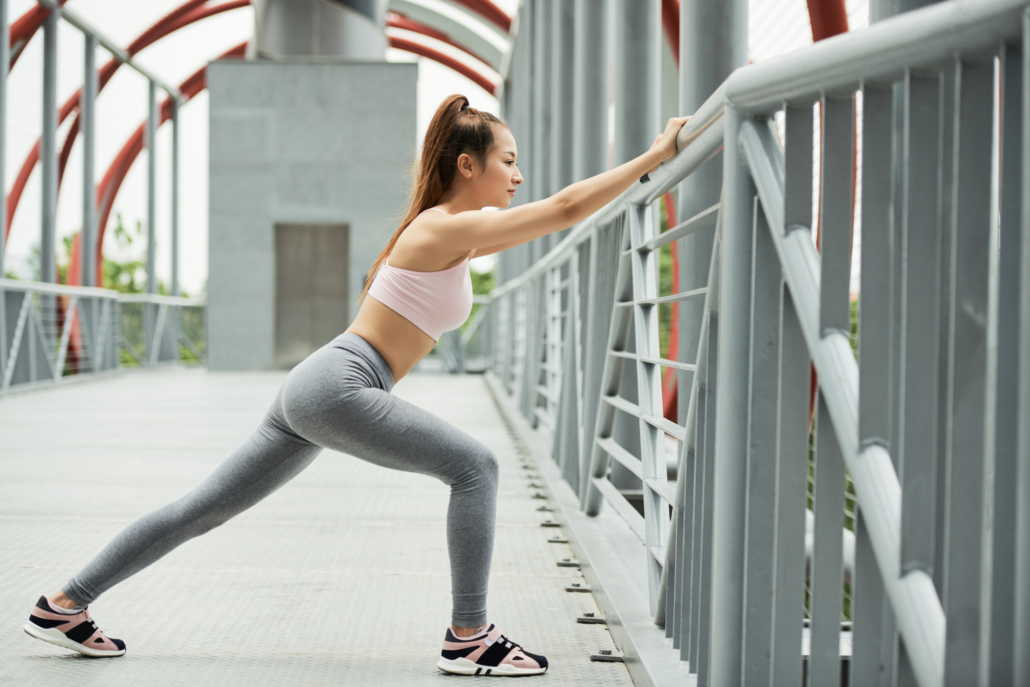
PEOPLE HAVE EXPERIENCED THE BENEFITS OF STRETCHING SINCE THE BEGINNING OF TIME. NOTHING RELIEVES A STIFF MUSCLE OR BACK BETTER THAN A GOOD STRETCH. MODERN SCIENCE HAS FORTUNATELY AFFORDED US THE KNOWLEDGE OF HOW, WHEN AND FOR HOW LONG IT IS BEST TO STRETCH.

THE EFFECTS OF STRETCHING
Stretching increases the length of your muscle, muscle fascia and tendons – thereby increasing flexibility and joint range of motion. Stretching also increases blood flow, lymph drainage and nerve activity in the muscle (these effects are usually measurable post-stretch.) This all leads to a healthy optimally firing muscle.

TYPES OF STRETCHING
There are many different types of stretching aimed to yield different results for different individuals’ needs. These can be broadly categorized into static, dynamic and ballistic stretching.

A) STATIC STRETCHING is the classic approach to stretching where the stretch is held for 30 to 60 seconds per muscle group. The aim here is to stretch and lengthen the muscle fibres, fascia and connective tissue. This stretch technique is however better performed post-workout and could in fact compromise muscle function if performed pre-workout. The reason for this is a decrease in muscle tension and an increase in length between resting muscle fibres. This affects the length-tension relationship of the muscle decreasing its excitability and muscle firing. Consider an elastic band which gets stretched for a long period of time and then released, it’s recoil is much slower than an elastic band which is stretched and released quickly. The stored elastic energy in a muscle behaves in a very similar fashion. This is why static stretches are not recommended pre-workout especially before explosive activities such as sprinting, jumping or throwing. Doing static stretching over a cold muscle can increase your risk of injury during the stretch and even during the work-out. These stretches are more beneficial post-workout to increase muscle and connective tissue length where necessary.

B) DYNAMIC STRETCHING is better suited to a pre-workout scenario. It is, however, also best done after light stimulation such as a 5 minute jog, row or spin to get the blood flowing through the desired muscle group. Dynamic stretching involves taking the body part gradually through its range of motion, increasing the range each time. This is done until maximum range of motion is reached. Each progressive stretch is held for a maximum of 2 to 3 seconds before relaxing, then repeating the cycle for a minute per targeted muscle group. Because the stretches are only held briefly the muscle is able to lengthen without losing the muscle tension or excitability. Muscle performance during the workout appears to be superior when warming up using the dynamic stretch technique, fatiguing less and maximizing output. This also invariably reduces the risk of injury.

C) BALLISTIC STRETCHING involves loading the muscle into the desired range of motion then bouncing or forcing it until full range is achieved. Although this technique does work quickly, the risk for injury during stretching is understandably far greater. I only recommend this style of stretching for experienced, elite athletes who are very in tune with their bodies’ stretch response.

THE DO NOT’S OF STRETCHING:
-Do not stretch over a totally cold muscle/first thing in the morning. After you get out of bed, first walk around and get active for a good 30min before stretching.
-Do not overstretch. Never stretch beyond the point of relative comfort.
-Do not stretch over a bruised/torn muscle. Always first have your injury assessed by a medical professional!
-Do not just stretch randomly over a chronic/relapsing injury-you could be doing more harm than good!
-Do not bounce your stretches, especially if you are doing a pre-workout stretching. Rather replace these with the dynamic stretches described above.

SO TO CONCLUDE, STRETCH BEFORE AND AFTER EXERCISE: Stick to dynamic stretching pre-workout and leave the static stretching for after! HOWEVER, REMEMBER NOT TO STRETCH RANDOMLY IN THE PRESENCE OF ACUTE OR CHRONIC INJURY! Get advised by your chiropractor regarding the stretches SPECIFIC FOR YOUR INDIVIDUAL INJURY. Written by Dr. E Greyling
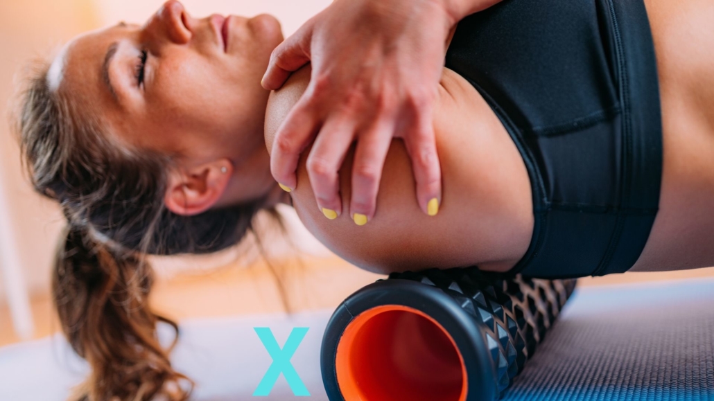
MUSCLE ROLLING HAS FAST BECOME ONE OF THE MOST POPULAR SELF-TREATMENTS IN THE WORLD. IT HAS BECOME THE GO-TO THERAPEUTIC TOOL FOR ATHLETES AND OFFICE WORKERS ALIKE. THERE ARE HOWEVER CONTRAINDICATIONS TO MUSCLE ROLLING, ESPECIALLY WHEN IT COMES TO THE SPINE.

The effects are very similar to massage but without the cost so it’s popularity is understandable!

The effects of muscle rolling include :
-increasing the flexibility of the muscle,
-increasing the blood flow in the muscle,
-increasing lymphatic drainage in the muscle,
-breaking down chronic muscle spasms and adhesions,
-loosening and stretching the fascia covering the muscle.

Despite the benefits, there are some serious contraindications to muscle rolling, DO NOT:
-roll over a haematoma (bruised muscle),
-roll over a muscle tear,
-roll over the calves or legs in the presence of painful swelling (rule out deep vein thrombosis).

MOST IMPORTANTLY FROM A CHIROPRACTIC PERSPECTIVE, DO NOT ROLL THE SPINE, ESPECIALLY NOT THE NECK!:
Unfortunately, the only techniques that can be used when self-rolling the spine involve almost your entire body weight. The spinal muscle layers are simply not thick enough to absorb all the pressure of your body weight on one point. This means an immense amount of pressure is being directed straight into the spinal joints.

If one considers the angulation of the spinal joints it’s easy to see that they are designed for axial loading, as in when standing – to carry and distribute the weight of the body down the spine. Rolling the spine unfortunately has a compressive or compacting effect on the spinal joints, which are not designed for that angle of sustained pressure. This action effectively compacts or blocks the joints causing joint dysfunction.

Often people who role their spines will attempt to self-manipulate, releasing a series of spinal clicks or cavitations. As discussed in the blog https://drchiro.co.za/is-clicking-your-own-joints-a-healthy-habit-what-is-the-difference-when-the-chiropractor-does-it/ANY joint can click and non-specific joint manipulation will only lead to joint HYPERMOBILITY (too much movement), inflammation and eventually arthritic change. Chiropractic manipulation SPECIFICALLY targets only the blocked and dysfunctional joints, and in the exact direction of the blockage, RESTORING NORMAL MOVEMENT.

Generally speaking, muscle rolling it is a safe practice yielding fantastic results! If, however, muscle rolling ever causes more pain post-treatment, or has any other adverse effect CONSULT A MEDICAL PROFESSIONAL – AND ALWAYS AVOID ROLLING THE SPINE!Written by DR. E Greyling

DO YOU SUFFER BACK OR NECK PAIN WHEN COUGHING OR SNEEZING. HERE IS WHAT COULD BE CAUSING THE PROBLEM.

When you cough or sneeze there is a sudden rise in your intra-spinal, or intra-thecal pressure. This increases the stress on already inflamed or impinged structures thus exacerbating the pain.

The same spinal pressure effect created when you cough or sneeze can be replicated clinically by performing the Valsalva manouvre. This can be done by bearing down hard as if going to the toilet or blowing out while holding your mouth and nose shut. Pain anywhere in the spine, especially when accompanied by shooting pain into the arms or legs, constitutes a positive sign for a focal inflammation or a space occupying lesion.

Usually a focal pain will indicate something as innocuous as a blocked or inflamed spinal facet joint (known as facet syndrome).
Nerve pain extending into the arms or legs would indicate a slightly more progressed spinal pathology- this is most often indicative of a prolapsed intervertebral disc (bulging disc) pinching a nerve. It may however also indicate a far more serious scenario such as a space occupying lesion pinching a nerve (spinal cysts, abscesses etc.) – these lesions are serious, but are also thankfully extremely rare.

Whether the cause of a positive Valsalva sign is innocuous or serious it needs to be further investigated. Always start more conservatively by visiting your Chiropractor to treat any spinal dysfunction and to rule out more serious pathology. Your Chiropractor will be the first to refer you to the appropriate specialist in the presence of any ‘red flags’ – this is however most times thankfully avoidable.

ALTHOUGH IT IS NOT THE SOLUTION – Relief from the actual pain experienced when coughing or sneezing can achieved by :

A) For cervical pain: Shrug the shoulders up completely and extend (hollow) the neck backwards slightly thereby increasing the cervical lordosis. This position braces the neck and cervical structures.
B) For thoracic pain: Shrug the shoulders backwards completely creating a pigeon chest effect while again extending (hollowing) the neck and lower back backwards. This position will brace the thoracic spine.
C) For lumbar pain: Shrug the shoulders back completely and extend (hollow) the thoracic and lumbar spine. Bend the knees placing both hands on the thighs deflecting all the pressure to the thighs. This position braces the lumbar spine and deflects the pressure to the thighs.

DO NOT EVER FLEX (ROUND) YOUR CERVICAL, THORACIC OR LUMBAR SPINE WHILE COUGHING OR SNEEZING IN THE PRESENCE OF A SPINAL INJURY. YOU WILL AGGRAVATE THE INJURY!

Don’t ignore the signs your body gives you. Pain is your body’s alarm system telling you that something is wrong… get it seen to! Written by DR. E Greyling

SITTING WITH YOUR LEGS CROSSED WILL DEFINITELY NOT CAUSE A MEDICAL EMERGENCY, BUT IT CERTAINLY CAN HAVE FAR REACHING NEGATIVE EFFECTS.

Sitting with your legs crossed will definitely not cause a medical emergency, but it certainly can have far reaching negative effects in individuals prone to certain conditions. Sitting with your legs crossed:


A) PRIMARILY causes structural compressive stresses to the calf muscles and all structures running through them- including blood vessels, lymph vessels and nerves.


*Prolonged compression to the calf muscles can cause activation of myofascial trigger points and local muscle spasm. This causes pain, loss of flexibility and calf muscle shortening-often leading to secondary conditions like achilles tendinitis and plantar fasciitis.


*Compression to any large muscle group such as the calf muscle causes a spike in blood pressure by forcing blood out of the muscle – this is obviously a concern in already hypertensive individuals.
The legs crossed posture also accentuates pooling of blood by cutting off blood flow leading to aggravation of varicose veins and potential thrombus (blood clot) formation.


*Compression to a nerve can result in a neuropraxia which is effectively a temporary break in the normal nerve transmission. This can result in pain, pins and needles, loss of sensation and weakness. We have all had a situation where our ‘leg goes to sleep’ when in a strange position -legs crossed is one of those positions and symptoms can last for weeks to months in more serious scenarios!

B) SECONDARILY sitting with the legs crossed creates functional torsional stresses to the knees, hips and pelvis:
*The torsional effect on the knee stresses the joint capsule and meniscus which can result in joint laxity or impingement scenarios.
*The torsional stresses around the hip joint also strains the joint capsule and creates hip muscle length/flexibility imbalances.
*The torsional or twisting stress at the pelvis causes a rotation pattern where one side of the pelvis tilts forwards and the other tilts backwards. This results in a functionally longer leg on the one side which causes compensatory imbalances all the way up the spine.

This does not mean you have to sit like a nun, by all means cross your legs at the ankles – JUST AVOID CROSSING YOUR LEGS AT THE KNEES. Please refer to the blog posted on November 21, 2019 https://drchiro.co.za/what-is-the-correct-office-posture-to-prevent-spinal-dysfunction/ for advice on how to sit correctly! Keep your fingers crossed, not your legs! Written by Dr.Etienne Greyling.